The electron microscope is a type of microscope that uses electrons to create an image of the target.
It has much higher magnification or resolving power than a normal light microscope.
Although modern electron microscopes can magnify objects up to two million times, they are still based upon Ruska's prototype and his correlation between wavelength and resolution.
Researchers use it to examine biological materials (such as microorganisms and cells), a variety of large molecules, medical biopsy samples, metals, and crystalline structures, and the characteristics of various surfaces.
Electron Microscopy is a technique for obtaining high-resolution images of biological and non-biological specimens. It is used for biomedical research for investigating the detailed structure of cells, tissues, macromolecular complexes, and organelles. The high resolution of the EM images results from the use of electrons having very short wavelengths as the source of the illuminating radiation. Electron microscopy is used in conjunction with a variety of ancillary techniques for answering specific questions. Key information on the structural basis of cell function and cell disease. An electron microscope is a type of microscope that uses electrons for creating an image of the target. It comes with very high magnification or resolving power than a normal light microscope. Despite the modern electron microscopes having the ability to magnify objects up to two million times, they are still based upon Ruska's prototype and his correlation between wavelength and resolution. For many laboratories, the electron microscope is an integral part. It is used by researchers for examining a variety of large molecules, biological materials, metals, and crystalline structures, the characteristics of various surfaces, and medical biopsy samples.
We have different types of Microscopes for different needs of the user.
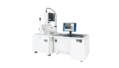
Scanning electron microscope (SEM)-Scanning Electron Microscopes (SEMs) are used across a number of industrial, commercial, and research applications. From cutting edge fabrication processes to forensic applications, there's a diverse range of practical applications for the modern SEM. This focuses the beam of electrons into a small spot which scans across the surface of a sample. The condenser lens assembles the electrons into a fine beam. The beam is focused onto the sample by the objective lens. The beam is caused to move in a rectangular X and Y direction by the deflection coils which produces a raster scan across the surface of the sample.
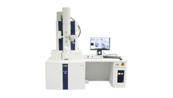
A TEM transmits the beam of electrons via a thin sample onto a screen or a detector. It has a large number of lenses. The amount of illumination which reaches the sample and control beam intensity or brightness is dependent on the condenser lenses. The objective lens focuses the beam of electrons onto the sample and applies a very marginal amount of magnification.

Scanning electron microscope (SEM)

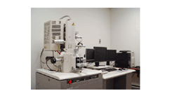
Field Emission Scanning Microscope (FESEM)

Transmission Electron Microscope (TEM)